Conduction System of the Heart
(A) Primary Pacemaker
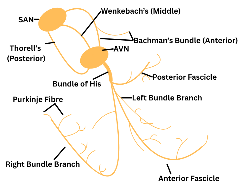
1. Sino-atrial Node (SAN): Primary pacemaker of the heart.
Cells of SAN have an intrinsic rate (frequency) of depolarization at approximately 70 beats/min.
(B) Secondary (latent) Pacemaker
2. Parts of the atrial myocardium: Cluster of atrial myocardial cells located around the crista terminalis, entrance of coronary sinus, IVC, around mitral & tricuspid valve, possess automaticity. These cells are not conduction cells per se; they are actually contractile cells that possess automaticity.
Intrinsic rate 60 beats/min.
3. Myocardium surrounding the Atrio-ventricular node (AVN): It is common misconception that the AVN possess automaticity because there is no compelling evidence for that. There is however, evidence that cell clusters surrounding the AVN possess automaticity. This automaticity still-despite what has just been stated be referred to as automaticity of the AVN in order to facilitate understanding.
Intrinsic rate - 40 beats/min.
4. The His-Purkinke Network: The bundle of His & the entire purkinje network possess automaticity.
Intrinsic rate of His bundle 20-40 beats/min, purkinje fibre 20-40 beats/min.
5. The ventricular myocardium does not possess automaticity
Propagation of impulses through conduction system
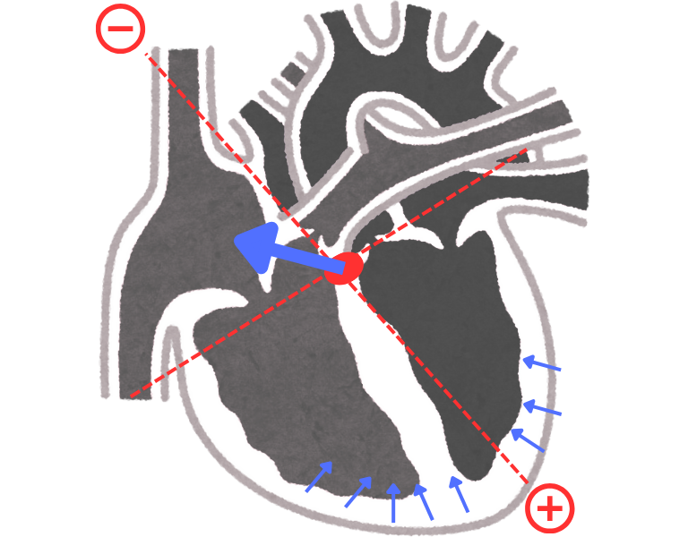
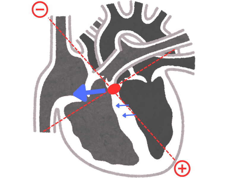
1. Atrial depolarization forms small vector because of thin myocardium & the vector is moderate with downward & leftward (Figure 2.). It shows positive deflection on ECG paper called, P wave.
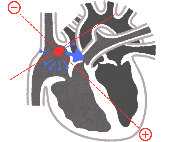
2. After complete atrial depolarization electrical vectors hits the annulus fibrosis (electrically insulates atria from ventricles) got cancelled (Figure 3.) because it is not a good conductor of current. Only window for electrical current to pass is AV node.
Electrical activity in AV node is very small that ECG machine can not pick up so during current is passing from AV node heart is electrically silent. This activity forms isoelectric line on ECG Paper called, PR segment
Note...
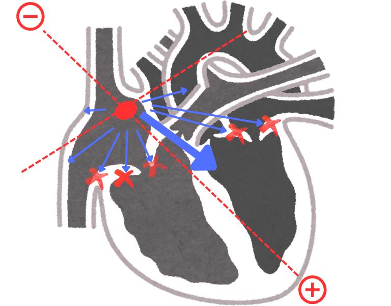
• AV node is specilized for slow conduction, so that atria got contraction properly & fills the ventricles.
• Purkinje fibers are very fast conductor that's why septum, ventricular walls & basal part of ventricles contracts simultaneously.
• Fibrous tissue make a seleev around bundle branches & fibrous covering made insulating layer on bundle brancehs.
| AV node | Purkinje fibre |
|---|---|
| More but small cells present hence more resistance due to cell membrane. | Less but large cells along the direction of current flow, less resistance. |
| Number of gap junctions between AV nodal cells are very very less. | Number of gap junctions between purkinje fibers are very much. |
| Ca2+ dependent depolarization present. (Ca2+ channels works slowely). | Na+ dependent depolarization present. (Na+ channels are fast). |
3. When ventricular septum is undergoing depolarization process depolarizing current moves from leftward & downward to rightward & upward (Figure 4.), vector is small & very fast. It shows negative deflection on ECG paper, called Q wave.
4. Ventricles depolarized simultaneously many vectors are produced they can be added together (Figure 5.). It produces positive deflection on ECG paper, called R wave.
Note...
• Purkinje fibers situated in subendocardial myocardium (deeper) hence depolarize first and then subpericardial myocardium.
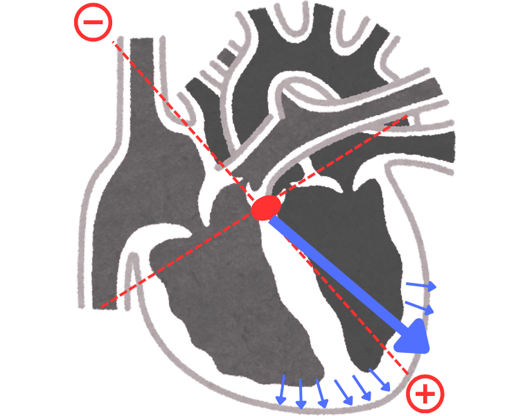
5. Depolarization toward basal ventricular part (Figure 6.) shows negative deflection on ECG paper called, S wave.
6. Complete depolarization (Figure 7.) on ventricles shows isoelectric line on ECG paper called, ST segment.
Note...
• Depolarization is fast process but repolarization is slow because Na+ channels are fast but K+ channels are slow.
• In depolarization first septum, then ventricular & then basal ventricular depolarization occurs hence there is 3 different identical waves are formed.
• In repolarization when septum starts its repolarization it has not complete yet major ventricular repolarization starts, it has not complete yet also basal ventricular repolarization starts.
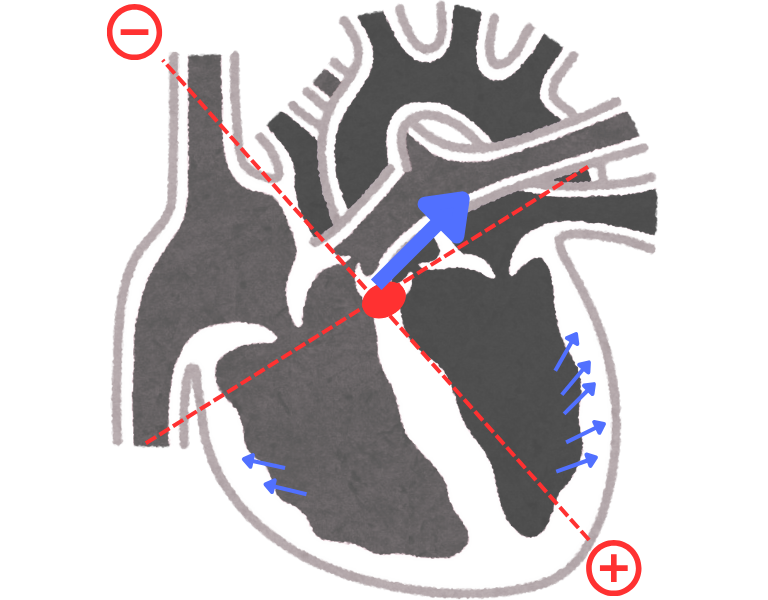
7. Repolarization current collapse or overlape. Only strongest vector deflects the needle in ECG machine & that vector is major ventricular repolarization. Because negative repolarization vector is toward negative lead the ECG deflection will be positive (Figure 8.). It shows positive deflection on paper called, T wave.